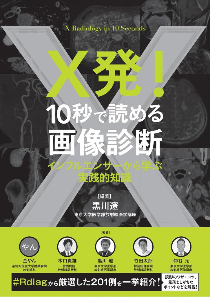
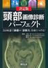
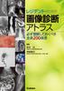
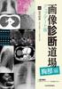
Quickly look at images and learn tips, tricks, and tricks for diagnostic imaging that will help you make future diagnoses!
Starting in April 2022, posts related to diagnostic imaging education on X (formerly Twitter) will be tagged with "#Rdiag." This book compiles important and easily misunderstood case studies, each explained on a separate page. The diagnostic areas are not limited to a single area, but cover a wide range, including the central nervous system, head and neck, chest, abdomen, bone and soft tissue/pelvis, the whole body, and more. This book contains 201 carefully selected cases, including images you've never seen before, rarely seen, images you've always wanted to see, and images you've seen but struggled to diagnose. It also includes information on commonly overlooked aspects of each case, as well as key points to consider when interpreting them. Another unique feature of this book is that many of the authors are winners of image interpretation competitions. Why not enjoy practicing diagnostic imaging with this book in hand and improve your own skills?
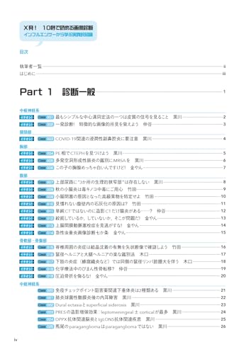
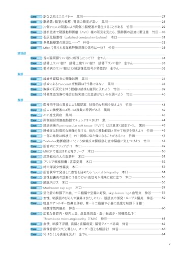
Table of Contents
Authors
Introduction
Part 1: General Diagnosis
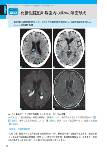
Part 2: Aunt Minnie (One-Shot Diagnosis)
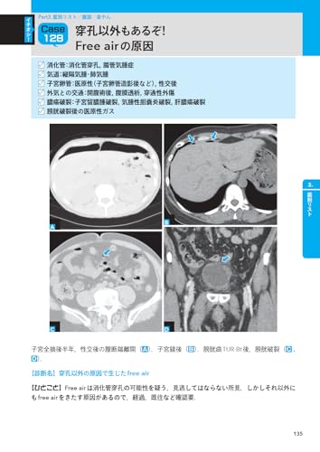
Part 3: Differential Diagnosis List
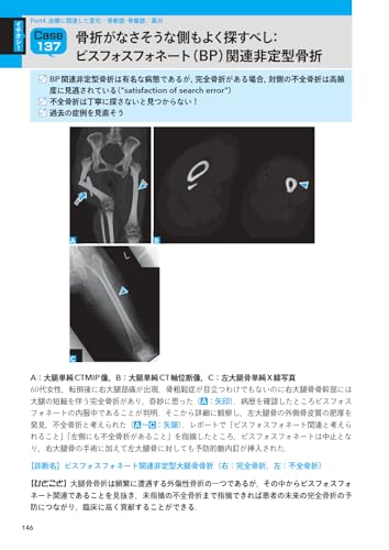
Part 4: Treatment-Related Changes
Part 5: Pitfalls
References
Author Profile
≫ Column 1: Keep Forgetting and Keep Learning. If Possible, Link It to an Anecdote
≫ Column 2: How to Make Learning Diagnostic Imaging "Fun"?
≫ Column 3: Make an Effort to Write About "What You Don't Know"
≫ Column 4: Studying with an Eye for Input-Output Balance
≫ Column 5: The Good Things I Did Before Becoming a Specialist, and What I Want to Continue Doing